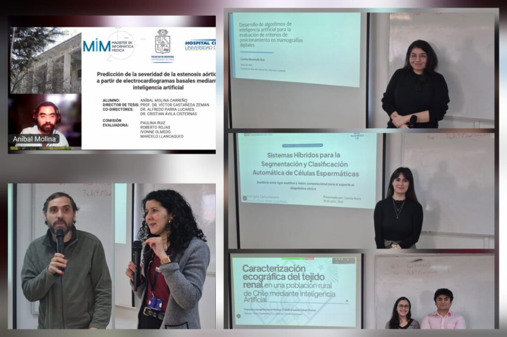
The AI and Innovation in Healthcare presentation day, organized by the Tech4MEDICS Lab, showcased research conducted by Medical Technology and Electrical Engineering students, highlighting the growing impact of artificial intelligence on clinical decision support. The event brought together faculty, researchers, students, and guests both in person and online.
Four thesis projects were presented, covering AI applications in medical imaging and biomedical signal analysis. Francisco Romero and Catalina Tobar introduced an AI-based framework for quantitative characterization of kidney tissue from ultrasound images, demonstrating robust segmentation performance and improved prediction of chronic kidney disease when combined with clinical variables. Camila Maire presented a hybrid system combining geometric methods and deep learning for automatic sperm cell segmentation and classification, achieving high accuracy while maintaining model interpretability. Carolina Bavestrello developed deep learning algorithms to automatically assess patient positioning quality in digital mammography, paving the way for automated quality control systems. Finally, Aníbal Molina presented AI models based on ResNet and XGBoost to predict aortic stenosis severity from routine electrocardiograms, showing promising generalization performance for non-invasive cardiovascular screening.
The event highlighted the high level of interdisciplinary research achieved by the students, integrating artificial intelligence, medical image processing, and deep learning to address clinically relevant challenges. As a milestone before their thesis defenses, these projects demonstrate the potential of AI-driven technologies to advance precision medicine and strengthen collaboration between engineering, medical technology, and health sciences.
Ultrasound-Based Characterization of Renal Tissue in a Rural Chilean Population Using Artificial Intelligence
Authors: Francisco Romero and Catalina Tobar
This work presented an artificial intelligence-based methodology for the quantitative characterization of renal tissue using ultrasound images obtained from the MAUCO cohort. The study included more than 760 patients and over 3,000 images, and developed an automated segmentation model that achieved Dice scores close to 0.9 for the main anatomical structures. Although the resulting biomarkers demonstrated stability under different image acquisition conditions, the findings indicate that, on their own, they are not yet sufficient to diagnose chronic kidney disease. However, when combined with clinical variables such as age and sex, they improve predictive performance, representing a complementary tool for future clinical applications.
Hybrid Systems for the Automatic Segmentation and Classification of Sperm Cells
Author: Camila Maire
This research proposed a hybrid system integrating geometric methods, analytical models, and deep neural networks for the automatic segmentation and classification of sperm cells. The algorithm achieved a semantic segmentation performance of approximately 95% and enabled the extraction of a highly interpretable set of morphological features through contour refinement techniques. Subsequently, these features were combined with a lightweight neural network based on attention mechanisms, achieving a classification accuracy above 84%, substantially outperforming traditional methods such as support vector machines. The study demonstrates the potential of combining explainable models with Deep Learning to support reproductive diagnostics.
Development of Artificial Intelligence Algorithms for the Assessment of Positioning Criteria in Digital Mammography
Author: Carolina Bavestrello
This research presented algorithms based on Deep Learning and geometric techniques to automatically assess positioning quality during digital mammography acquisition, considering both craniocaudal and mediolateral oblique views. The developed models achieved excellent segmentation performance for relevant anatomical structures, with an average Dice score of 0.902 and an average IoU of 0.832. These results represent an important step toward automated quality control systems that could contribute to improving the standardization and reliability of mammographic examinations.
Prediction of Aortic Stenosis Severity from Baseline Electrocardiograms Using Artificial Intelligence
Author: Anibal Molina
This research presented the development of artificial intelligence models aimed at estimating the severity of aortic stenosis from baseline electrocardiograms, exploring a non-invasive approach to support the early diagnosis of this cardiovascular disease. The study demonstrates the potential of deep learning to extract clinically relevant information from routinely acquired electrocardiographic signals, contributing to the development of clinical decision-support tools and strengthening AI-assisted diagnosis in cardiology. ResNet and XGBoost models were evaluated using two approaches: a cascade model and a multiclass model. External validation demonstrated promising generalization potential, although further confirmation in larger and more diverse cohorts is required.